Figure 2.

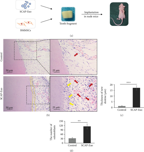
Exosomes from the stem cells of the apical papilla (SCAP-Exo) promoted the regeneration of the dentine-pulp complex in vivo. (a) Schematic diagram of the animal experiment: SCAP-Exo, bone marrow mesenchymal stem cells (BMMSCs), and gelatine sponges as scaffolds were inserted into the canal of the tooth fragments, while the control group was treated with the same preparation without SCAP-Exo. The tooth fragments were then implanted into the nude mice. (b) HE staining showed a newly continuous layer of dentine (dotted line), odontoblast-like cells with overt polarised morphology (yellow arrow), and enhanced vascular formation (red arrow) in the experimental group. (c, d) The thickness of the new dentine and the number of odontoblasts were higher in the SCAP-Exo group than that in the control group (∗∗P < 0.01, ∗∗∗P < 0.001, n = 10). Error bars indicate means ± SD.
