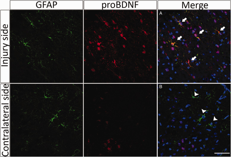
Figure 4.
proBDNF Is Induced in Astrocytes After TBI. Adult mice were subjected to CCI and perfused 3 days after injury. Representative sections through the injury site 3 days after CCI show increased proBDNF labeling (red), some of which colocalizes with GFAP (green) adjacent to the area of tissue damage. In contrast, sections through the contralateral side 3 days after the injury show little expression of proBDNF. (A) Arrows indicate colocalization of proBDNF and GFAP-positive cells. (B) Arrowheads indicate GFAP-positive cells that do not express proBDNF. Scale bar = 50 µm.
GFAP = glial fibrillary acidic protein; proBDNF = pro-brain-derived neurotrophic factor.

