Figure 6.
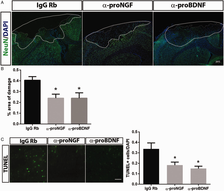
Neutralizing Antibodies to proNGF and proBDNF Provide Neuroprotection. (A) Mice were infused intranasally with anti-proNGF, anti-proBDNF, or control IgG immediately after the CCI. At 3 days of recovery, sections were stained for NeuN and counterstained with DAPI to reveal the area of damage. (B) The area of total damage comprised of the area of tissue loss and the penumbra (dotted line), where the density of DAPI and NeuN staining was reduced. The percentage of the total area of damage (relative to the contralateral hemisphere) was significantly reduced by the antiproneurotrophin antibodies. Scale bar = 200 µm. (C) Representative images of TUNEL staining in the penumbra showed fewer apoptotic cells in the mice that received anti-proNGF or anti-proBDNF. Scale bar = 50 µm. Data were collected from 3 to 4 animals per group. Graphs depict the means ± SEM. Asterisks indicate significance by one-way analysis of variance followed by Tukey’s post hoc analysis with p < .05.
IgG = immunoglobulin G; proBDNF = pro-brain-derived neurotrophic factor; proNGF = pro-nerve growth factor; DAPI = 4′,6′-diamidino-2-phenylindole; TUNEL = terminal deoxynucleotidyl transferase deoxyuridine triphosphate nick end labeling.

