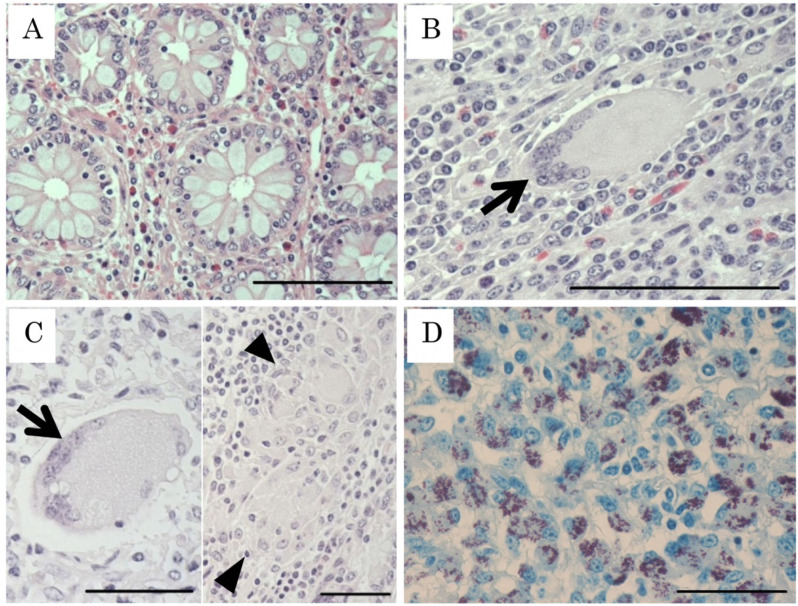
Fig. 1.
Histopathological classification of Johne’s disease lesions. (A) Non-lesional type (N type). Normal structure of crypta is observed. Hematoxylin Eosin (HE); Bar=100 µm. (B) Tuberculoid type (T type). Lymphocyte and macrophage infiltration in the apex of the villus and the formation of multinucleated giant cell (arrow) are observed. HE; Bar=100 µm. (C) Mixed type (T/L type). A multinucleated giant cell and accumulation of epithelioid cells (arrow head) are observed. HE; Bar=50 µm. (D) Lepromatous type (L type). Epithelioid cells containing Mycobacterium avium subsp. paratuberculosis (MAP) infiltrate in the ileal submucosa and accumulate predominantly between the lamina propria and muscular layer. Ziehl-Neelsen staining; Bar=50 µm.

