
Fig. 5.
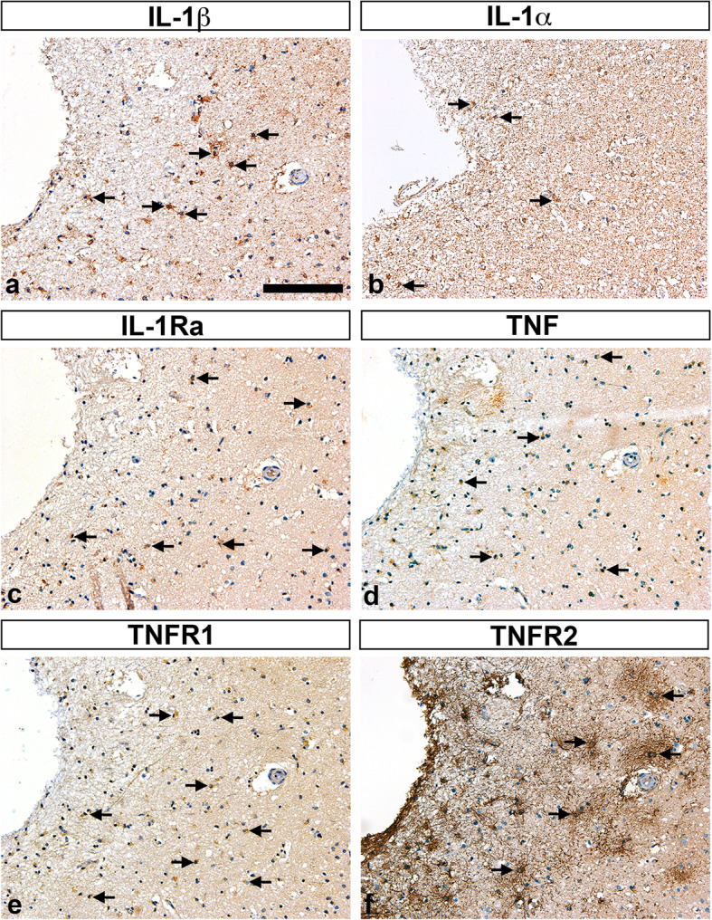
Characterization of IL-1, TNF, and TNF receptors TNFR1, and TNFR2 in post-mortem tissue sections. a-f Immunohistochemical staining of parallel tissue sections from a > 7-day-old infarct for cytokines IL-1β (arrows in a), IL-1α (arrows in b), IL-1Ra (arrows in c), and TNF (arrows in d), and for TNFR1 (arrows in e), and TNFR2 (arrows in f). IL-1α, interleukin-1alpha; IL-1β, interleukin-1beta; IL-1Ra, interleukin-1 receptor antagonist; TNF, tumor necrosis factor; TNFR1, tumor necrosis factor receptor 1; TNFR2, tumor necrosis factor receptor 2. Scale bar = 40 μm