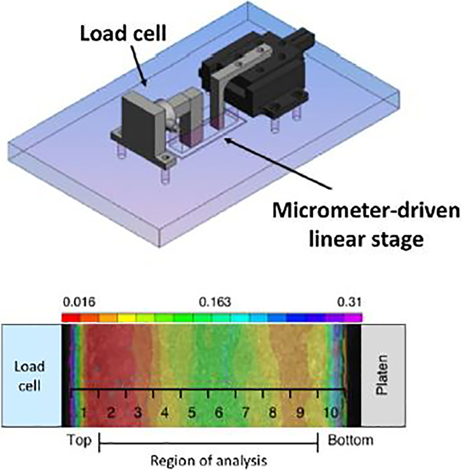
Fig. 1.

Schematic of custom compression device mounted on an inverted fluorescent microscope and heat map of strain mapped to ten distinct regions of analysis from the superficial to deep zone. Adapted from Farrell et al. (2012), used with permission.
