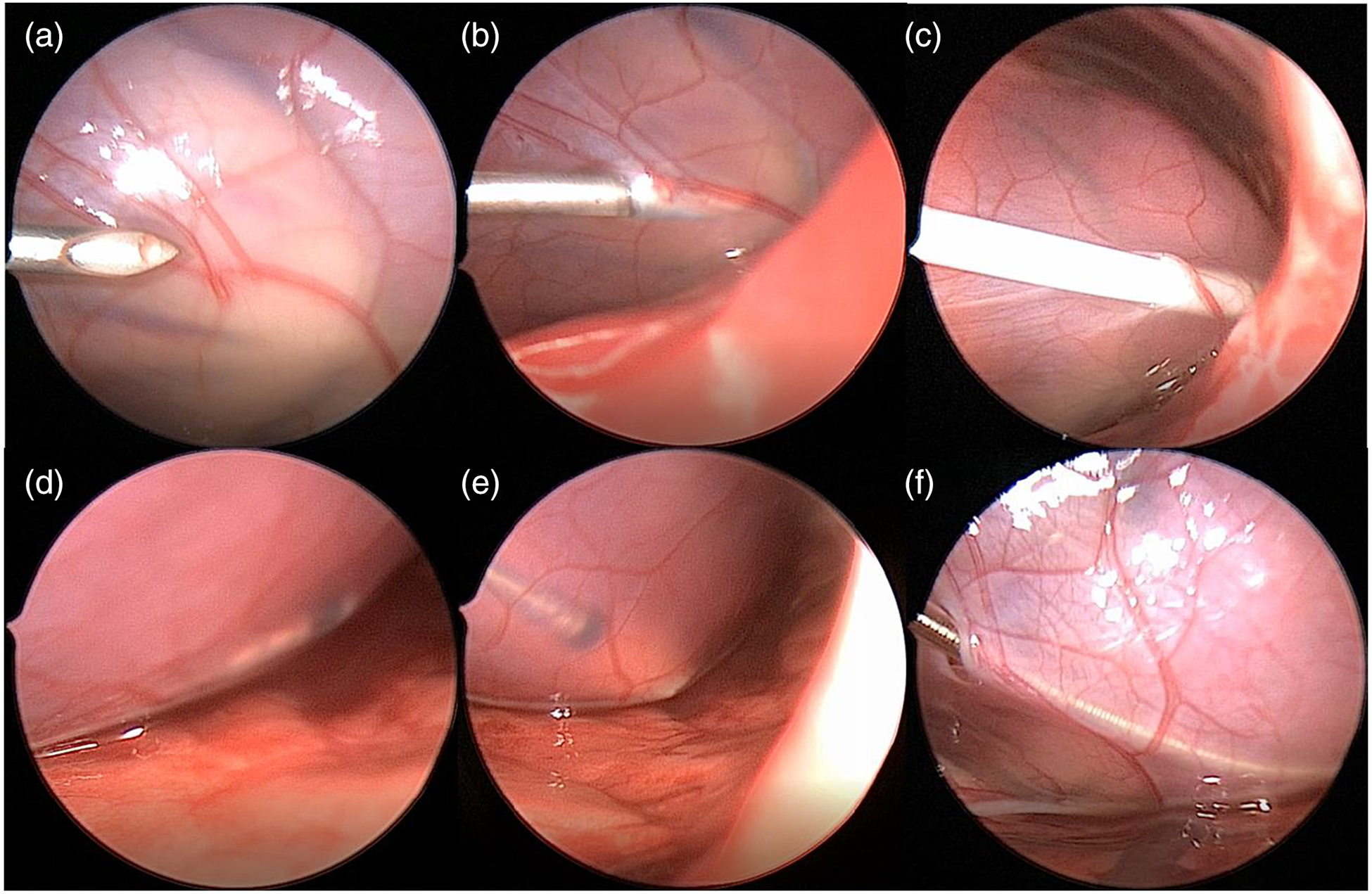
Fig. 3.

Procedural workflow as seen by thoracoscopic visualization a needle visualization, b needle insertion into pericardial space, c successful sheath insertion into pericardial space, d distal pacemaker lead placement within pericardial space, e zoomed up image of pacemaker lead tip, f completed procedure with lead in place and small defect remaining in pericardium
