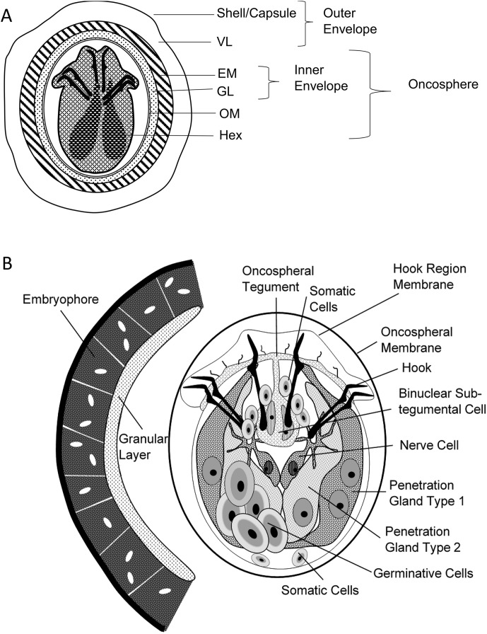
Figure 2A and B.
General description of the egg and oncosphere of Echinococcus spp, according to Jabbar et al., 2010 [27]. (A) Schematic diagram of an oncosphere illustrating the structure and bilateral symmetry in the pattern of hooks and cellular organization of the hexacanth embryo. VL: vitelline layer; EM: embryophore; GL: granular layer; OM: oncospheral membrane; Hex: hexacanth embryo. (B) Cellular organization of the oncosphere. Oncospheres are approximately 25 × 30 μm.

