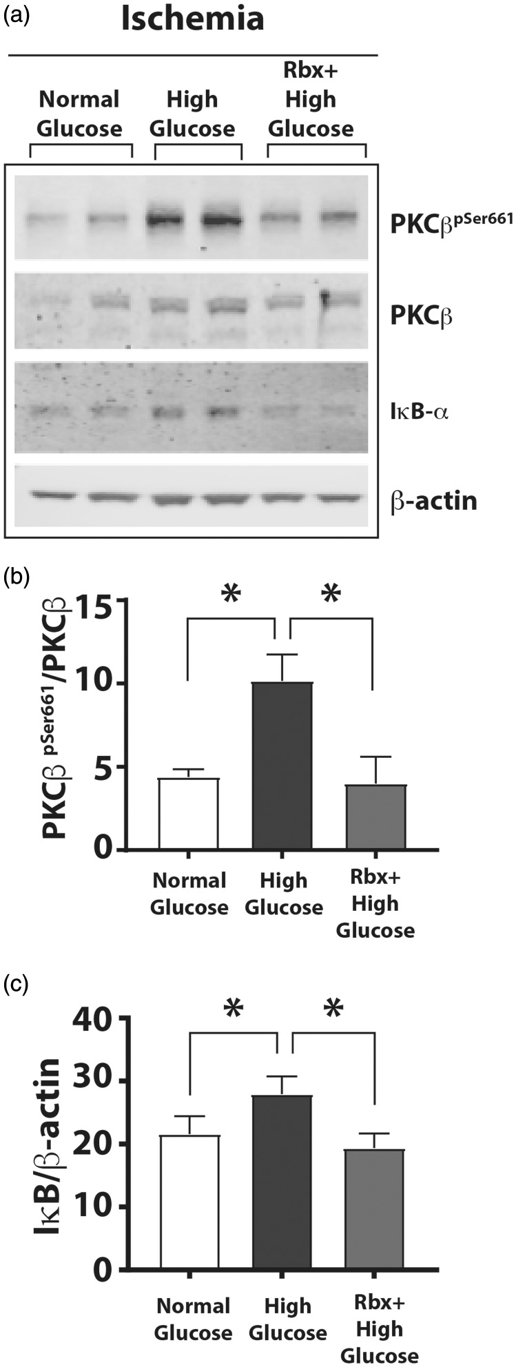
Figure 6.
A PKCβ inhibitor Rbx decreased phosphorylation of Ser661 residue of PKCβ and increased IκBα degradation. HUVEC were grown in normal, high D-glucose, or high D-glucose + Rbx with ischemia for 24 h. (a) Western blots of HUVEC grown in different glucose concentrations with ischemia. (b) Autophosphorylated PKCβpSer661band showed greater intensity in cell lysates with high D-glucose that was decreased in the cells grown in the high D-glucose + Rbx (20 nM). (c) The IκBα levels (IκBα/β-actin) in the corresponding samples were increased in high D-glucose but were significantly reduced in the cells treated with Rbx. Asterisks denote P < 0.05, and n.s. denotes not significant; n= 4 to 6 per group.
PKCβ: protein kinase C beta; Rbx: ruboxistaurin.

