
Figure 1. TCF21 suppresses SMC lineage marker expression.
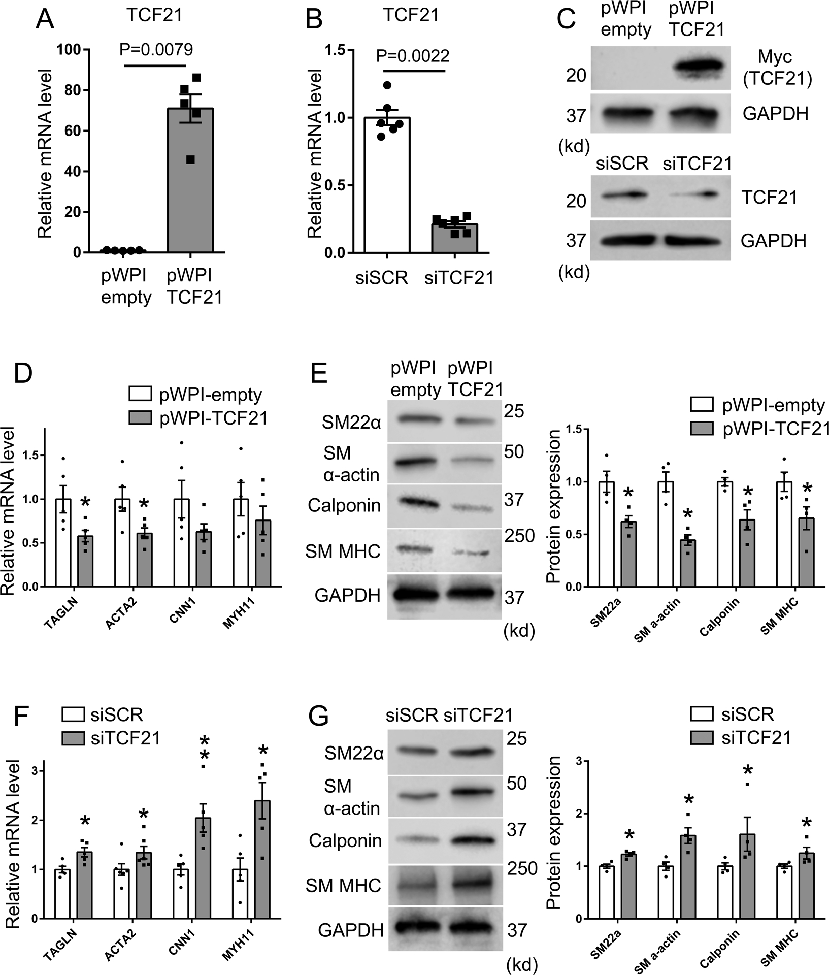
A-C) For overexpression or knockdown of TCF21, HCASMC were transfected with TCF21 overexpressing lentivirus (pWPI-TCF21) or siTCF21, respectively. mRNA and protein expression of TCF21 was evaluated by qPCR or western blot. D) HCASMC were treated with TCF21 overexpressing lentivirus (pWPI-TCF21) or empty control lentivirus (pWPI-empty) for 24 hours, and mRNA was evaluated by qPCR for expression of SMC markers TAGLN, ACTA2, CNN1, MYH11 (n=5), (p* values, TAGLN; 0.0317, ACTA2; 0.0317). E) HCASMC were treated with TCF21 overexpressing lentivirus (pWPI-TCF21) or empty lentivirus (pWPI-empty) and protein expression of SMC markers evaluated by western blot. Band intensity of SMC markers was quantified by image J software and normalized to GAPDH (n=4) (all p* values; 0.0286). F) HCASMC underwent TCF21 knockdown with transfection of siTCF21 or control siSCR regulatory RNAs and SMC marker expression evaluated by qPCR (n=5–6) (p* values, TAGLN; 0.0317, ACTA2; 0.026, MYH11; 0.0317; p** value, CNN1; 0.0079). G) HCASMC underwent TCF21 knockdown with transfection of siTCF21 or control siSCR regulatory RNAs and protein expression of SMC markers evaluated by western blot. Band intensity was quantified by image J software and normalized to GAPDH (n=4) (all p* values; 0.0286). Data analyzed by Mann-Whitney U test.