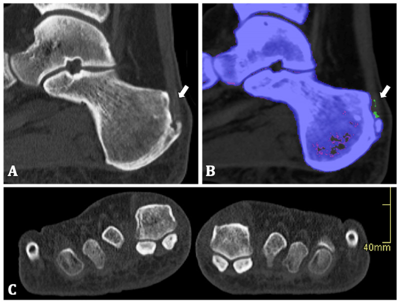
Figure 2.

50-year-old man. (A) Sagittal native CT image of the right calcaneum. Calcaneal enthesophyte and several linear areas of high density in the Achilles enthesis are shown (arrow). (B) Colour-coding identifies the linear densities as uric acid crystals (arrow). (C) Coronal native CT image shows there were no erosions of the MTP-I.
