Figure 4.

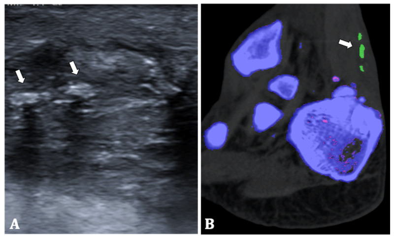
70-year-old man. (A) Ultrasound of the right Achilles tendon shows tendon thickening and multiple hyperechoic nodules (arrows). (B) Colour-coded DECT image shows MSU crystal deposition in the Achilles tendon (arrow).

70-year-old man. (A) Ultrasound of the right Achilles tendon shows tendon thickening and multiple hyperechoic nodules (arrows). (B) Colour-coded DECT image shows MSU crystal deposition in the Achilles tendon (arrow).