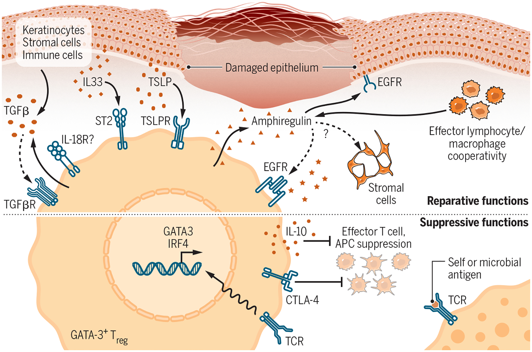
Figure 3-. Reparative and Suppressive Effector Mechanisms of Type 2-Polarized Tregs.

Skin bears a high proportion of type 2-polarized Tregs programmed by Th2-associated transcription factors such as GATA-3 and IRF4 that both confer tissue-reparative functions. Top: GATA-3+ Tregs in skin express receptors for alarmins such as TSLP, IL-33, and possibly IL-18 that are released upon tissue damage, enabling them to sense local injury. IL-18 and IL-33 can both stimulate Treg production of the reparative cytokine amphiregulin independent of TCR stimulation. Amphiregulin (AREG) drives keratinocyte proliferation and may promote regeneration of stromal populations in wounded skin. Skin Tregs additionally express both TGF-β and TGFβR, although the Treg-specific function of each is not entirely clear. Many of these reparative functions are shared by effector lymphocyte and macrophage populations. Bottom: In addition to conferring direct tissue-reparative function to Tregs, GATA-3 and IRF4 also bolster adaptive Treg functions in part by stabilizing FoxP3 expression. These “classical” Treg functions are linked to TCR stimulation and include production of the regulatory cytokine IL-10 and suppression of costimulation on antigen presenting cells by Treg-expressed CTLA-4. Solid arrows, known interactions; Dotted arrows, likely interactions based on data from other tissues and contexts.
