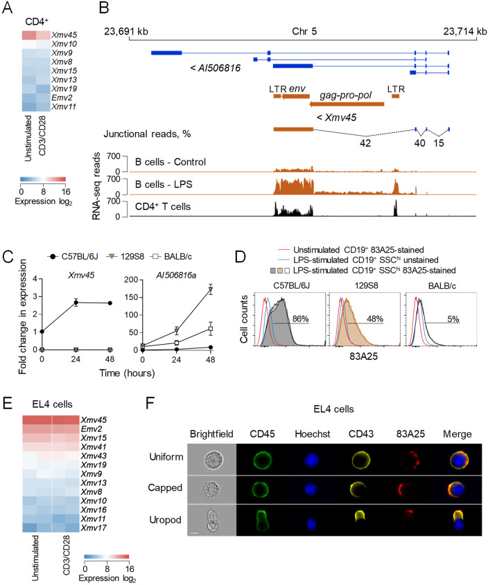
Fig 1. An endogenous MLV env is an immune stimulation-responsive gene in B cells and is expressed by primary lymphocytes and EL4 thymoma cells.
(A) Heatmap of endogenous MLV expression in resting and CD3/CD28-stimulated CD4+ T cells assessed by RNA-seq. (B) Genomic location of Xmv45 and RNA-seq read mapping in unstimulated and LPS-stimulated B cells and in unstimulated CD4+ T cells. (C) Splenocytes of C57BL/6J, 129S8 or BALB/c mice were in vitro stimulated with LPS for 24 or 48 hours and the expression of Xmv45 (left) and AI506816a (right) was assessed by qRT-PCR. Expression levels following LPS stimulation were compared to basal expression levels in unstimulated C57BL/6J splenocytes. (D) Flow cytometric analysis of MLV envelope on the surface of SSChi B cells from C57BL/6J, 129S8 or BALB/c mice following in vitro stimulation with LPS for 48 hours compared with unstimulated, but stained B cells and with stimulated, but 83A25-unstained B cells. Two mice per strain are shown. (E) Heatmap of endogenous MLV expression in resting and CD3/CD28-stimulated EL4 cells assessed by RNA-seq. Two biological replicates are shown. (F) MLV envelope localisation in non-polarised and polarised EL4 cells. EL4 cells were labelled with anti-CD45, anti-CD43 for 30 minutes and 83A25 for 15 minutes at 37°C, counterstained with Hoechst and imaged by IS. Scale bar = 7 μm.

