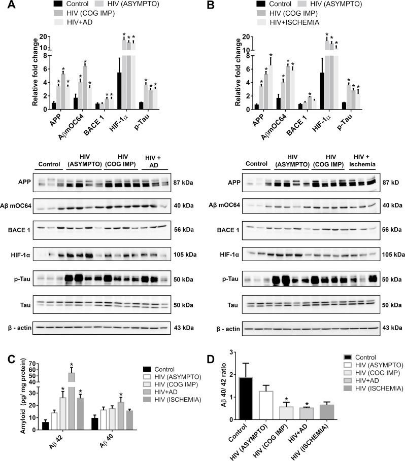
Fig 1. Amyloidopathy and neurofibrillary tangles in the FCs of individuals infected with HIV.
(A) Representative western blots showing the expression of APP, Aβ mOC64, BACE1, HIF-1α, p-Tau, and Tau in the FC of control (uninfected, n = 3), patients infected with HIV with asymptomatic [HIV (ASYMPTO), n = 4], cognitive impairment [HIV (COG IMP), n = 4], those with neurofibrillar pathology (HIV + AD, n = 3), (B) as well as with cerebral ischemia (HIV + Ischemia, n = 3). β-actin was used as an internal control. (C) ELISA showing the protein levels of Aβ42 and 40 and (D) graphical representation of Aβ40/42 ratio. Data are presented as mean ± SEM. One-way ANOVA followed by Bonferroni post hoc test was performed, *P < 0.05 versus control. The data underlying this figure may be found in S1 Data. AD, Alzheimer Disease; APP, amyloid precursor protein; Aβ, amyloid beta; BACE1, β-site cleaving enzyme; FC, frontal cortex; HIF-1α, hypoxia-inducible factor.

