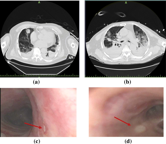
Fig. 1.
Lung CT scans and bronchoscopy findings of patients with COVID-2019 and IPA. Patient 1, 90 years, lung CT scan showed nodules with cavities in the middle lobe of the right lung and consolidation bilateral lower lobes with a small amount of pleural effusion (a); bronchoscopy finding showed small ulcer in the right wall of the trachea (c). Patient 2, 74 years, lung CT scan showed consolidation bilateral lower lobes with multiple irregular cavities in the middle (b); bronchoscopy finding showed right middle bronchial ulcer (d). Red arrow indicated the positions of bronchial ulcers

