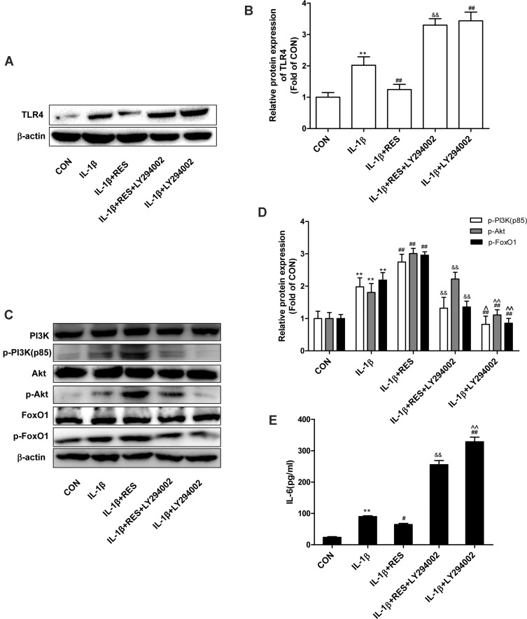
Figure 4.
PI3K/Akt regulated TLR4 and FoxO1 expression. (A) Serum-starved (0.5% FBS) SW1353 cells were pretreated with LY294002 (25 μg/mL) for 1 h and then exposed to IL-1β (10 ng/mL) with or without resveratrol (50 μM) for 24 h. TLR4 expression and were determined by Western blotting analysis. (B) The levels of TLR4 were normalized with β-actin. The results for Western blot were expressed as folds of CON. (C) After SW1353 cells pretreated with LY294002 were exposed to IL-1β (10 ng/mL) in the absence or presence of resveratrol (50 μM) for 30 min, p-PI3K, p-Akt, and p-FoxO1 expression were determined by Western blotting analysis. (D) The levels of p-PI3K, p-Akt and p-FoxO1 were normalized with their respective total PI3K, Akt, FoxO1 levels. The results for Western blot were expressed as folds of CON. (E) IL-6 concentrations in the culture supernatants incubated for 24 h were determined by ELISA. All data above were expressed as the mean ± SD of three independent experiments. **P <0.01 versus the CON group, #P <0.05, ##P <0.01 versus the IL-1β group, &&P <0.01 versus the IL-1β + RES group, ^P <0.05, ^^P <0.01 versus the IL-1β + RES + LY294002 group.

