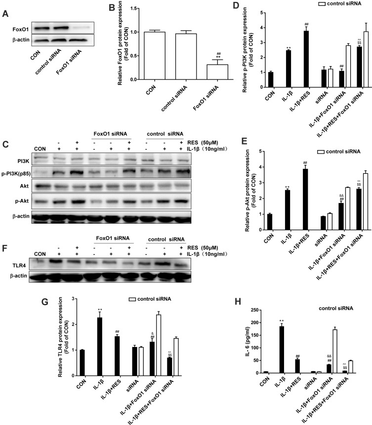
Figure 5.
FoxO1-knockdown decreased p-PI3K, p-Akt and TLR4 protein expression in IL-1β-treated SW1353 cell. (A) SW1353 cells were transfected with FoxO1 siRNA (50 nM) or control siRNA for 48 h, FoxO1 expression was analyzed by Western blot. (B) The levels of FoxO1 were normalized with β-actin. The results for Western blot were expressed as folds of CON. **P <0.01, versus the CON, ##P <0.01 versus control siRNA. (C), (F) After SW1353 cells were transfected with FoxO1 siRNA for 48 h as described above, cells were exposed to 10 ng/mL IL-1β with or without 50 μM RES for 30 min or 24 h, p-PI3K, p-Akt, and TLR4 expression was analyzed by Western blot. (D–E), (G) The levels of p-PI3K, p-Akt, and TLR4 were normalized with their respective total PI3K, Akt or β-actin levels. The results for Western blot were expressed as folds of CON. (H) IL-6 concentrations in the culture supernatants were assessed by ELISA. All data above were expressed as the mean ± SD of three independent experiments. **P <0.01 versus the CON, ##P <0.01 versus the IL-1β, $$P <0.01 versus IL-1β + RES, &P <0.05, &&P <0.01 versus siRNA, ^^P <0.01 versus siRNA + IL-1β.

