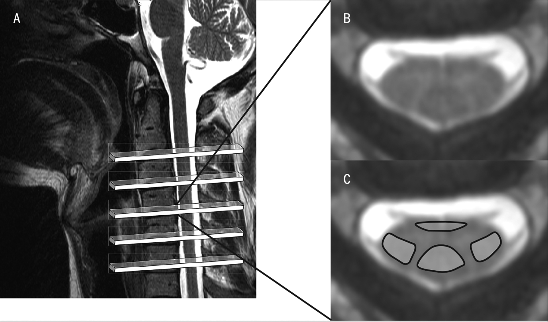
FIGURE 4.

(A) A T2-weighted magnetic resonance image was used to plan the axial slices for MT- and non—MT-weighted acquisition, spanning the C2–7 area of the spinal cord. (B) Magnetization transfer image corresponding to the region caudal to C4. (C) Non-MT image in the same region, with anatomically defined regions of interest over the ventromedial and dorsal columns of the spinal cord. These images are used to measure the MT ratio, quantifying anomalies in white-matter pathways. Abbreviation: MT, magnetization transfer.
