Figure 2.

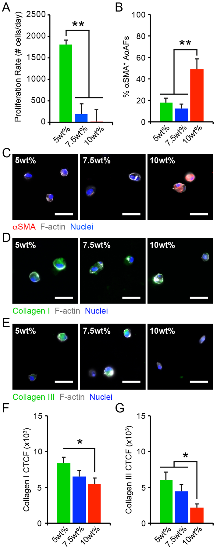
(A) The proliferation rate of AoAFs encapsulated in 5wt%, 7.5wt%, and 10wt% hydrogels significantly decreased with increasing weight percent over 7 days. (B-C) The percentage of αSMA+ AoAFs was significantly increased in 10wt% hydrogels compared to softer 5wt% and 7.5wt% substrates after 1 day, as confirmed by immunocytochemistry. Blue: Hoechst 33258 (nuclei), Grey: F-actin, Red: αSMA. Scale bar = 25 μm. (D-E) AoAFs produced (D) collagen I or (E) collagen III, detected via immunocytochemistry, following encapsulation in 5wt%, 7.5wt%, and 10wt% hydrogels for 1 day. Blue: Hoechst 33258 (nuclei), Grey: F-actin, Green: collagen I or collagen III. Scale bar = 25 μm. (F-G) Expression of (F) collagen I and (G) collagen III expression, measured by corrected total cell fluorescence, decreased with increasing weight percent. Data represented as the mean ± SEM, where in A: n = 3 biological replicates per condition and in C & F-G, n = 20 cells from 3 images from each of 3 hydrogels per condition. For A, C, & F-G: a one-way ANOVA, followed by a Tukey HSD post hoc test, was used to detect statistical significance, *p<0.05, **p<0.01.
