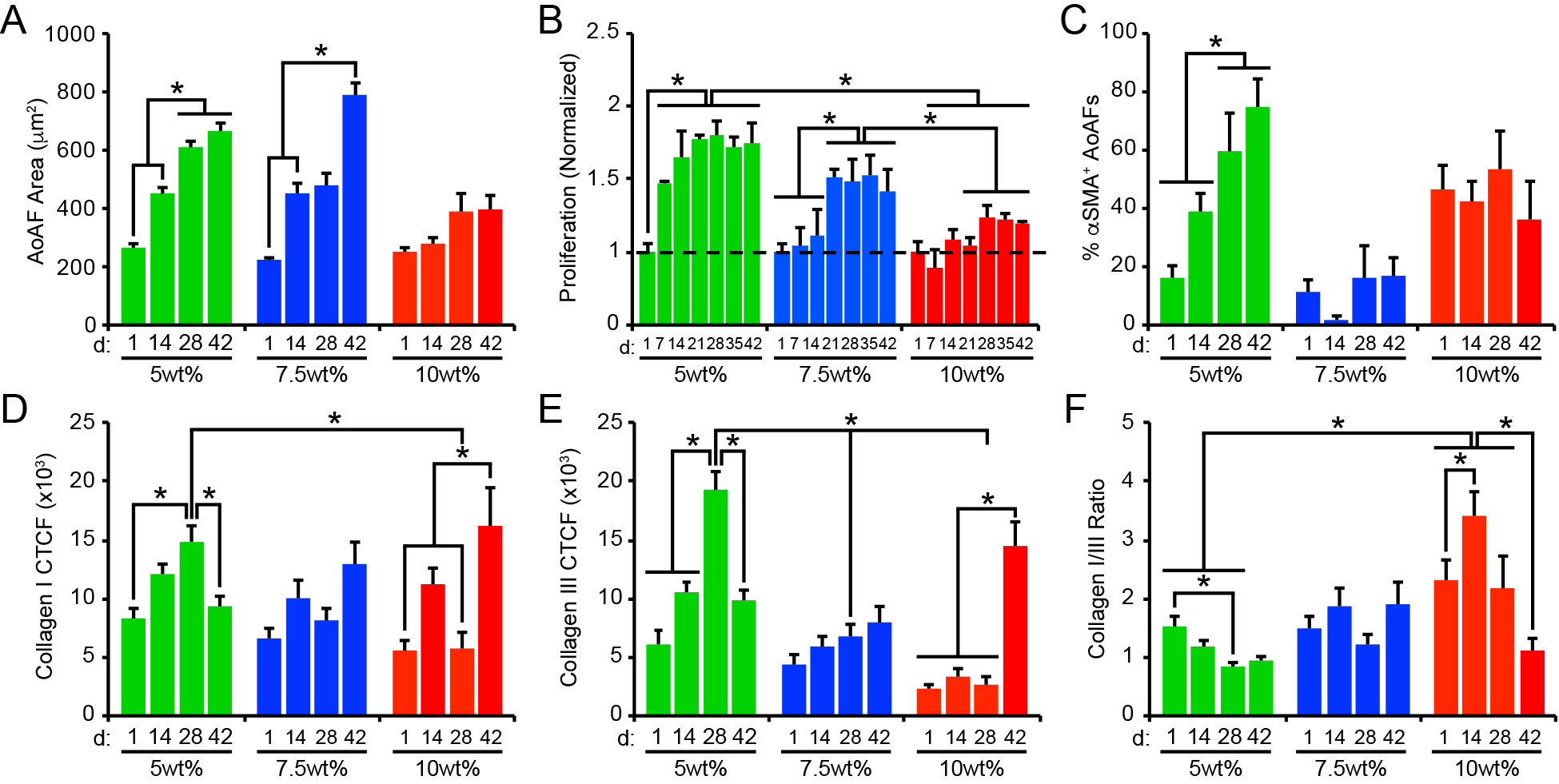
Figure 4.

(A) Cell area increased over time in 5wt% and 7.5wt% hydrogels, but not in 10wt% substrates. (B) Similarly, AoAF proliferation increased in 5wt% and 7.5wt% hydrogels over time. Data are mean DNA content relative to DNA content in hydrogels after 1 day of culture (dashed line) ± SEM, n = 3 biological replicates per condition. (C) The percentage of αSMA` AoAFs was significantly increased in 5wt% hydrogels over time, but alterations were not observed in 7.5wt% and 10wt% hydrogels. Expression of (D) collagen I and (E) collagen III, measured via corrected total cell fluorescence, by AoAFs encapsulated in 5wt%, 7.5wt%, and 10wt% hydrogels was altered over time. (F) Ratio of collagen I to collagen III, determined from total collagen signal obtained from the corrected total cell fluorescence, in AoAF-laden 5wt%, 7.5wt%, and 10wt% hydrogels over time. In A-F, data are represented as mean ± SEM, n = 3 biological replicates per condition. A two-way ANOVA, followed by a Tukey HSD post hoc test, was used to detect statistical significance, *p<0.05, **p<0.01.
