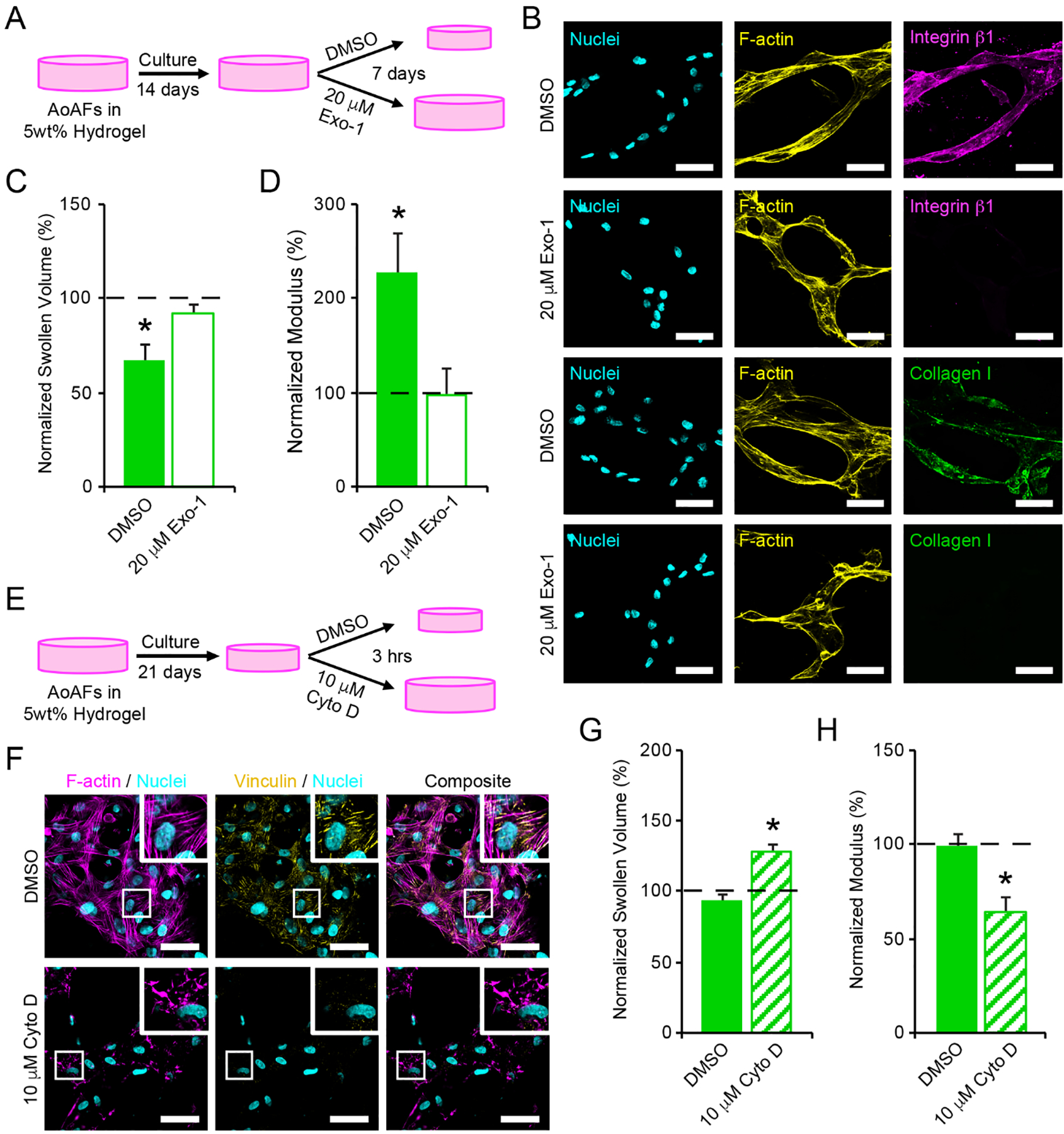
Figure 5.

(A) To investigate whether cell interactions with the hydrogel network were responsible for contraction of 5wt% hydrogels, encapsulated AoAFs were cultured for 14 days prior to an additional 7 days of culture in medium supplemented with or without 20 μM Exo-1. (B) Treatment with 20 μM Exo-1 for 7 days decreased integrin β1 and collagen I expression by AoAFs in 5wt% hydrogels. Cyan: Hoechst 33258 (nuclei), yellow: F-actin, magenta: integrin β1, green: collagen I. Scale bar = 50 μm. (C-D) Hydrogels treated with 20 μM Exo-1 for 7 days did not exhibit (C) decreased swollen volume or (D) increased hydrogel modulus. (E) To assess the importance of cytoskeletal tension in maintaining hydrogel contraction, encapsulated AoAFs were cultured in 5wt% hydrogels for 21 days and then exposed to medium with and without 10 μM Cyto D for 3 hrs. (F) Treatment with 10 μM Cyto D for 3 hrs decreased F-actin polymerization and focal adhesion formation, as indicated by vinculin, by AoAFs in 5wt% hydrogels. Cyan: Hoechst 33258 (nuclei), magenta: F-actin, yellow: vinculin. Scale bar = 50 μm. (G-H) Following exposure to 10 μM Cyto D, (G) swollen volume of 5wt% hydrogels increased and (H) hydrogel modulus decreased. Data are mean ± SEM, n = 3 biological replicates per condition. For C-D & G-H: A repeated measures student’s t-test, followed by a Tukey’s HSD post hoc test, was used to detect statistical significance from day 14 measurements (dotted line) and from DMSO treated controls. In C-D: *p<0.05 for DMSO control relative to pre-treatment measurements on day 14 and to Exo-1-treated scaffolds on day 21. In G-H: *p<0.05 for Cyto D-treated scaffolds relative to pre-treatment measurements (dotted line) and to DMSO treated controls.
