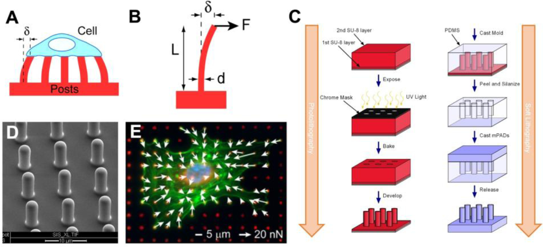
Figure 4.

Cellular forces can be measured using microposts. (A) Cells deflect the microposts as they contract and the amount of force can be calculated from the deflection of the posts (δ). (B) Each post is a cantilever beam of length (L) and diameter (d) that deflects in proportion to the force applied at its tip (F). (C) Using photolithography techniques, a master structure of the microposts is made using SU-8 photoresist on a silicon wafer by exposing it to UV light through a chrome mask, baking it to crosslink the SU-8, and then using a solvent to develop the final master structure by removing the unexposed SU-8. Next, soft lithography techniques are used create a negative mold of the microposts by casting them in PDMS, then fluorosilanizing the mold and using it to cast the final PDMS structure. (D) An array of PDMS microposts imaged by scanning electron microscopy. (E) Cellular forces are measured by quantifying the defection of the posts which have been labeled with a fluorescent dye (red). White arrows indicate direction and magnitude of the forces, green is F-actin staining, and blue marks the nucleus. C is adapted with permission.[51] Copyright 2007, Elsevier. E is adapted with permission.[52] Copyright 2012, Elsevier.
