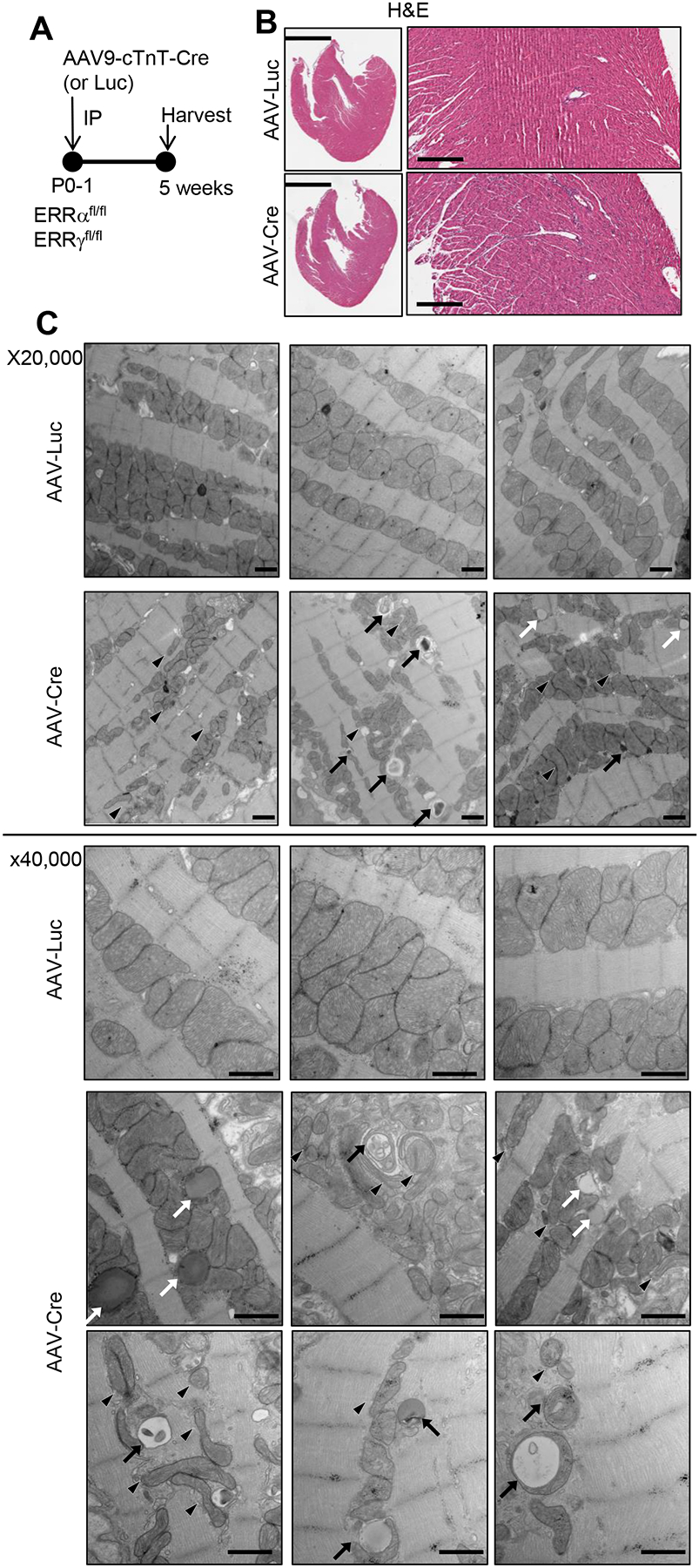
Figure 2. Inducible cardiac ERRϖ/γ deficiency results in abnormalities in mitochondrial density and structure.

(A) Schematic showing the experimental timeline. AAV-Cre or AAV-Luc (control) was administered to P1 ERRα/γflox/flox pups at a dose that does not cause cardiac dysfunction as described in the text. (B) Representative H&E images of male AAV-Luc or AAV-Cre-injected 5 week-old LV. Scale bars represent 2 mm in lower magnification and 300 mm in higher magnification. (C) Representative electron microscope images of LV wall of the male or female postnatal ERR KD hearts (x20,000 and 40,000) magnification. Arrows indicate structurally abnormal mitochondria including engulfed organelles indicative of mitophagy (black arrows), and elongated or fragmented (black arrowheads). White arrows indicate droplet-like structures that may represent lipid. Scale bars represent 800 nm.
