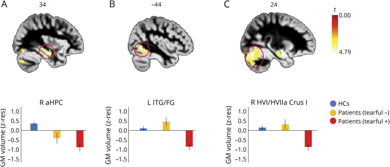
Figure 2. Structural abnormalities in patients with pathologic tearfulness.
Results of whole-brain voxel-based morphometry (VBM) on modulated gray matter (GM) (reflecting GM volume). Contrast: healthy controls (HCs) and patients without pathologic tearfulness > patients with pathologic tearfulness; between-subjects nuisance regressors: age, sex, total intracranial volume (TIV), and study (Memory and Amnesia Project [MAP], Oxford Project To Investigate Memory and Ageing [OPTIMA]). (A) Right anterior hippocampus: kE = 19, p family-wise error-corrected (FWE) = 0.037; peak voxel: t = 4.79; x = 34, y = −12, z = −17. (B) Left fusiform gyrus/posterior portion of inferior temporal gyrus: kE = 17; p FWE = 0.038; peak voxel: t = 4.79; x = −44, y = −62, z = −5. (C) Right cerebellar hemispheric lobules VI/VIIa Crus I: kE = 23; p FWE = 0.042; peak voxel: t = 4.76; x = 24, y = −75, z = −18; clusters are displayed here at p < 0.001 (unc) for display purposes, and survive FWE correction (p < 0.05) at peak-voxel level over p < 0.001 (unc) (minimum cluster volume: kE > 10). The cerebellar cluster also survived correction for nonstationary smoothness and cluster size (p-FWE < 0.05). Clusters are overlaid here on a diffeomorphic anatomical registration through exponentiated lie algebra GM template in Montreal Neurological Institute space (sagittal sections presented); heat bar represents t values; bar graphs display the average GM volume of each of those 3 clusters for the 3 different groups; error bars represent +1/−1 SEM. aHPC = anterior hippocampus; FG = fusiform gyrus; ITG = inferior temporal gyrus; kE = cluster size (number of voxels); R, L = right, left (hemisphere); z-res = mean values residualized against age, sex, study (MAP, OPTIMA), and TIV across participants.

