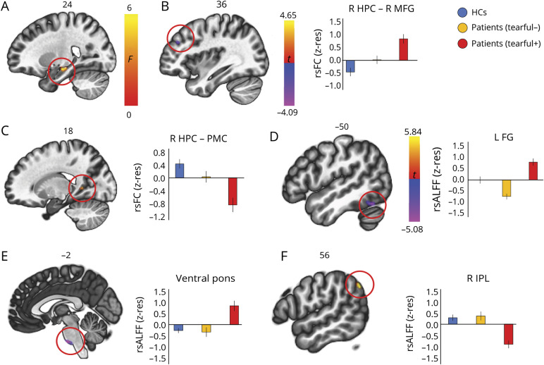
Figure 3. Functional abnormalities in patients with pathologic tearfulness.
(A) A connectome–multivariate pattern analysis demonstrated that patients with pathologic tearfulness, as compared with the rest of the patients and healthy controls (HCs), showed abnormal resting-state functional connectivity (rsFC) between a region in the right hippocampal (HPC) head and body and the rest of the brain; right hippocampal head and body: 24 −16 −14, kE = 194; p family-wise error-corrected (FWE) (cluster-level) = 0.001. (B-C) Patients with pathologic tearfulness showed (B) increased rsFC of the right hippocampus with the right middle frontal gyrus (MFG) (x = 36, y = 40, z = 30; kE = 105, p FWE = 0.03, t = −4.09), and (C) reduced rsFC of the right hippocampus with the posteromedial cortex (PMC) (peak voxel: t = 4.65; x = 18, y = −50, z = 6; kE = 98, p FWE = 0.04), extending to the right lingual gyrus. (D–F) Patients with pathologic tearfulness showed aberrantly increased resting-state amplitude of low frequency fluctuations (rsALFF) as compared with both the rest of the patients and healthy controls in (D) the left fusiform gyrus (kE = 69; peak voxel: t = −4.72; x = −50, y = −60, z = −20), and (E) the ventral pons (kE = 74; peak voxel: t = −5.08; x = −2, y = −20, z = −44; p FWE = 0.04), and reduced rsALFF in (F) the right inferior parietal lobule (peak voxel: t = 5.84; x = 56, y = −62, z = 40; kE = 112; p FWE = 0.004). All clusters survive FWE correction (p < 0.05) for cluster size over an uncorrected individual voxel threshold of p < 0.001. Error bars represent ± 1 SEM. FG = fusiform gyrus; IPL = inferior parietal lobule; kE = cluster size; R, L = right, left (hemisphere); z-res = mean rsFC and rsALFF values are residualized against age and sex across participants.

