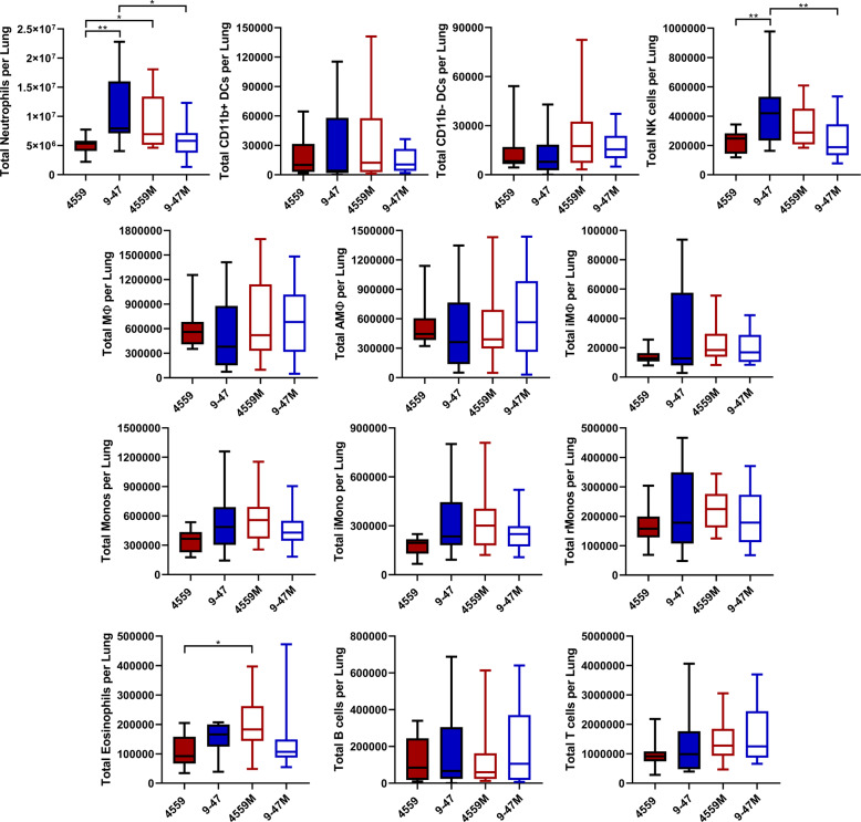
Fig. 6. Quantification of immune cell subsets in murine lungs 6 h post infection.
Groups of 8 mice per strain were challenged with either 9–47-Ear, 4559-Blood, 9–47M or 4559M. Single cell lung suspensions were prepared and stained with antibodies against various surface markers (Supplementary Table 4) then analyzed by flow cytometry. Populations enumerated include: NK natural killer cells, iMono neutrophils, eosinophils, inflammatory monocytes, rMono resident monocytes, AMФ alveolar macrophages, iMФ interstitial macrophages, CD11b−DC CD11b-negative dendritic cells, CD11b+DC CD11b-positive dendritic cells, T cells and B cells. Graphs shown represent pooled data from two independent experiments. All quantitative data are presented as mean ± S.E.M (n = 16 for each group), analyzed by one-way ANOVA (*p < 0.05; **p < 0.01).

