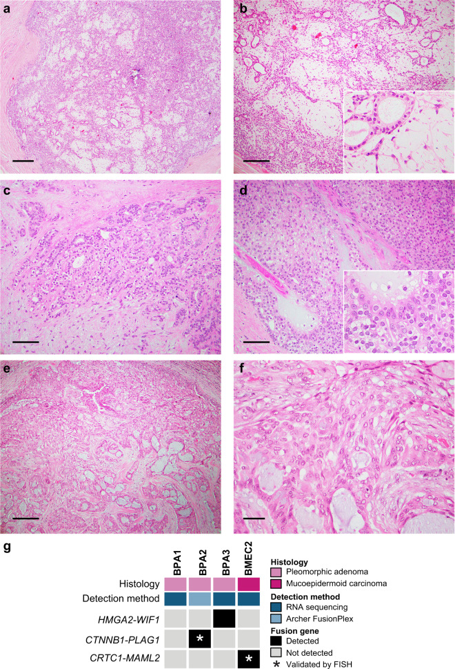
Fig. 1. Histologic features of and fusion genes in breast pleomorphic adenomas and a mucoepidermoid carcinoma.
a–d Representative photomicrographs of hematoxylin and eosin (H&E)-stained sections of breast pleomorphic adenomas (PAs) included in this study. Breast PAs were well-circumscribed lesions (a), composed of epithelial and myoepithelial cells in a myxochondroid stroma (b, c). Focal areas with solid growth were also identified (d). e, f Representative photomicrographs of H&E-stained sections of the breast mucoepidermoid carcinoma (MEC) displaying squamous, mucinous, and intermediate cells (e), and absence of atypia or necrosis (f). g Heatmap depicting fusion genes identified in breast pleomorphic adenomas (BPAs; n=3) and a breast mucoepidermoid carcinoma (BMEC2; n=1). Cases are shown in columns and fusion genes in rows. Tumor type and detection method are shown (top). Scale bars, 200μm (a), 100μm (b, e), 50μm (c, d), and 20μm (f). FISH fluorescence in situ hybridization.

