Fig. 2.

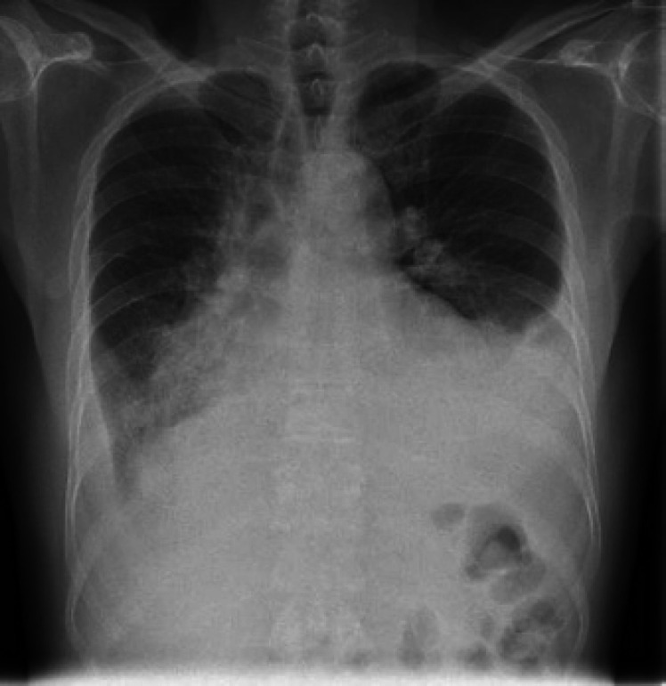
PA erect chest X-ray shows bilateral lower lobe collapse and consolidation with pleural effusion, more noted on the left side.

PA erect chest X-ray shows bilateral lower lobe collapse and consolidation with pleural effusion, more noted on the left side.