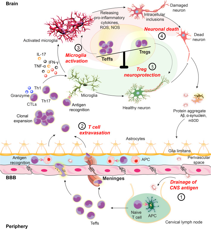
Fig. 2.
Teff activities promote neuroinflammation. In neurodegenerative disorders, CNS antigens including Aβ, α-syn, mSOD and MBP provoke microglia immune responses leading to neuroinflammatory cascade in affected brain regions. (1) Self-antigen or misfolded proteins are generated from damaged neuronal cells. The neural antigens drain to the peripheral lymphoid nodes by meningeal lymphatic vessels where they are taken up by local APCs including macrophages and DCs. Natural self-antigens are presented to peripheral T cells in MHCII dependent manner. (2) Naïve T cells, upon recognition of cognate antigen, differentiate into antigen-specific Teffs. Reactive microglia secrete cytokine-chemokine milieu to upregulate cell adhesion molecules (CAM) by blood-brain barrier (BBB) endothelial cells, opening the gate for peripheral primed T cells. Teffs (Th1 and Th17) with upregulated integrins and CAM ligands readily cross the BBB. Teffs also cross the blood-CSF barrier through choroid plexus meninges. After extravasation into the brain, Teffs are reactivated upon recognition of cognate antigen on common CNS APCs. These include perivascular macrophages (PVMs), choroid plexus and meningeal macrophages and DCs and parenchymal microglia. (3, 4) Activated Teffs secrete pro-inflammatory and neurotoxic mediators to polarize microglia to a higher activation state, producing pro-inflammatory cytokines and reactive oxygen and nitrogen species which further perpetuate the inflammatory cascade to induce neurotoxicity. (5) Tregs maintain immune tolerance by suppressing effector immune responses. Naïve T cells can also differentiate into Tregs upon recognition of cognate antigen on peripheral APCs in secondary lymphoid tissues. Differentiated Tregs exert neuroprotective responses through multiple mechanisms. The inflammatory immune responses observed in neurodegenerative disorders are the outcome of Teff-Treg imbalance with upregulated Teff responses

