Fig. 4.
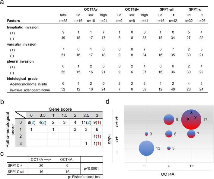
The OCT4/SPP1 axis in early-stage LUAD. a Summary of pathohistological factors, as well as OCT4- and SPP1-transcript expression in tumours from 58 LUAD patients. Values represent the number of patients harbouring tumours with designated characteristics. b Relationship between gene expression in the OCT4A/SPP1 axis and pathohistological scores. OCT4A scores: not detected (0), low levels (1), and high levels (2); SPP1 scores: not detected (0), SPP1-all detected (0.5), and both SPP1-all and SPP1C detected (1). Gene scores represent the total scores of OCT4A and SPP1. Criteria for evaluation of their expression levels was shown in Fig. S3. The pathohistological score is indicated as a total of each pathological score: Lymphatic invasion (Ly) (+): 1; vascular (Ve) (+): 1; and pleural invasion (pl) (+): 1. Blue numbers indicate cases of adenocarcinoma in situ, and red numbers indicate relapse cases. c Correlation between OCT4A and SPP1C expression in tumour tissues. The X-axis indicates SPP1C-expression levels (detected or undetected), and the Y-axis indicates OCT4A-expression levels (high and low levels or undetected). d Scatter plot showing the relationship between gene expression related to the OCT4A/SPP1 axis and levels of pathohistological risk. Each scatter point is represented by the following: an X-coordinate for OCT4A-expression levels (−: undetected; Low: detected at low levels; and High: detected at high levels), a Y-coordinate for SPP1-all and/or SPP1C expression (−: undetected; a: detection of SPP1-all detected; a + c: detection of SPP1-all and SPP1C); plot size: case number (each number indicates the number of cases); and colour [each colour represents pathohistological risk (blue: negative; and red: positive)]. Three black “X”s indicate that all three patients relapsed or had distant metastases during the observation period (case nos. 16, 59, and 62; Table S5)

