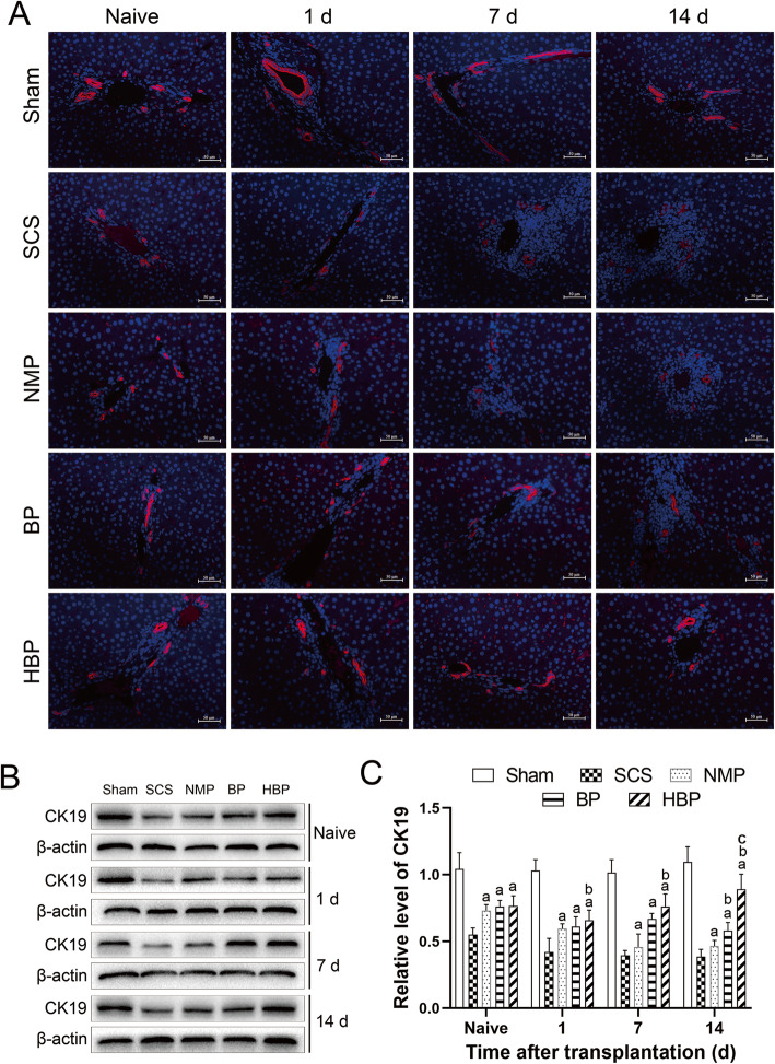
Fig. 6.
Expression of CK19 in biliary epithelial cells of the transplanted liver. A Immunofluorescence staining for CK19 (× 200, red tag) showed that the percentage of CK19-positive cells among biliary epithelial cells in the HBP group was significantly higher than that in the other groups. B Expression of CK19 in the transplanted liver analyzed by western blotting. C Relative expression of the CK19 protein (CK19/β-actin) showed that the relative expression of CK19 in the HBP group was significantly higher than that in the other groups

