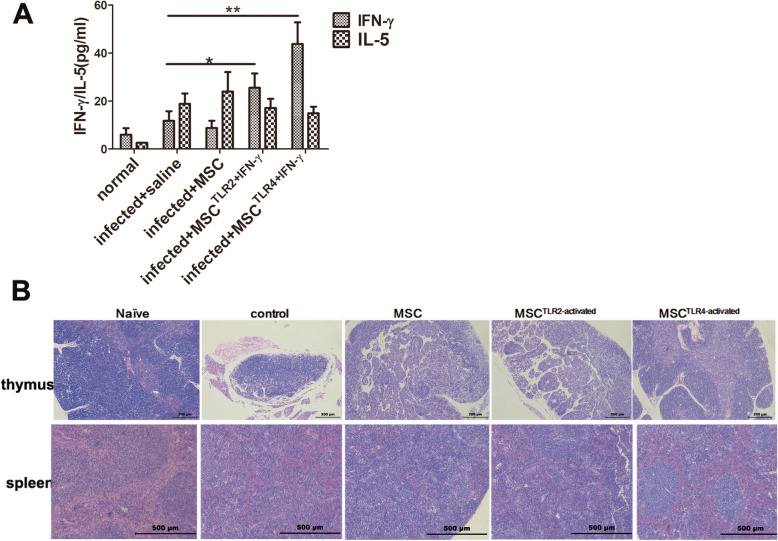
Fig. 4.
The TLR4+IFN-γ-activated MSC group had the highest levels of IFN-γ in the serum with an improved structure of the thymus and spleen, and TLR2-treated MSC and unpretreated MSC showed a mild increase of IFN-γ and no significant increase of IFN-γ, respectively. To measure systemic IFN-γ and IL-5 protein levels, the serum was taken from 8-week-infected mice and used in capture ELISAs (a), and histological change was evaluated by HE staining (b). Data are presented as the means ± SEM from 7 to 10 mice in each group. *P < 0.05; **P < 0.01

