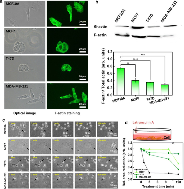
Fig. 2.
F-actin distribution, amounts, and depolymerization. a Distribution of F-actin in four different cells, observed by optical (left column) and fluorescence (right column) microscopy. b Western blot analysis of soluble actin (G-actin) and insoluble actin (F-actin). The ratio of F-actin to total actin (G- and F-actin). c Morphological change in cells induced by the LatA treatment. The same position on the cell was indicated with an arrow to compare morphological changes according to the treatment time. d Relative cell area reduction as a function of LatA treatment time. Each value represents mean ± SEM (*p ≤ 0.05, **p ≤ 0.01, ***p ≤ 0.001, ****p ≤ 0.0001)

