Fig. 1.
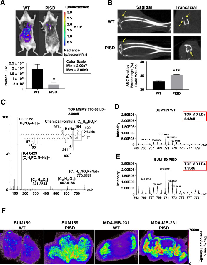
PISD reduces metastasis of triple-negative breast cancer cells. a Bioluminescence images of mice injected via the left cardiac ventricle with MDA-MB-231-WT control or PISD expressing cells depict reduced metastatic burden for mice injected with cells that stably express PISD. Graph shows mean whole-body photon flux + SEM for mice that survived to the experimental endpoint (n ≥ 7). *p < 0.05. b Sagittal and transaxial computed tomography (CT) images of tibias obtained 40 days after femoral artery injection of MDA-MB-231-WT or PISD cells. Images demonstrate reduced osteolytic bone metastases (arrows) in mice with MDA-MB-231-PISD cells. Graph displays mean values for area-under-the-curve for remaining bone volume in proximal tibia + SEM (n = 6). ***p < 0.0001. c MALDI-MS/MS spectrum of the sodiated ion (770.55 m/z) of 18:0 PE from cell pellets. d, e Graphs show mass spectrometry time-of-flight (TOF) signal intensity (red boxes) of 18:0 PE (770.55 m/z) in cell pellets from SUM159-WT (d) and PISD cells (e). f MALDI imaging mass spectrometry images of the sodiated ion of 18:0 PE detected at m/z 770.552 in orthotopic breast tumors from SUM159- or MDA-MB-231-WT or PISD cells. Scale bar = 5 mm

