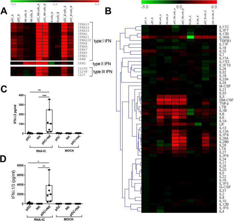
Fig. 1.
NK and B cells enhance the type III IFN production in pDCs stimulated with RNA-IC. a, b Relative signal intensity (log2fold change) of mRNA expression in RNA-IC-stimulated, vs mock-stimulated, cells from two healthy blood donors (a and b) after 6 h. Green indicates relative downregulation, black neutral, and red relative upregulation of gene expression. Protein levels of c IFN-λ2 and d IFN-λ1/3 in supernatants after 20-h stimulation. Boxplots show medians with interquartile range (seven donors, three independent experiments). Friedman’s test. *p < 0.05, **p < 0.01, ***p < 0.001

