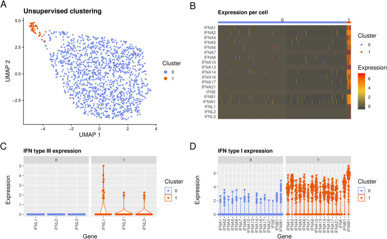
Fig. 4.
Type I and type III IFN expression in pDCs on the single-cell level. a Results from single-cell RNA sequencing illustrated by unsupervised clustering of 1413 healthy blood donor (n = 2) pDCs by non-linear two-dimensional Uniform Manifold Approximation and Projection (UMAP) embedding. Cells were stimulated with RNA-IC, IL-3, and IFN-α2b. Cluster “0” (blue) and cluster “1” (orange). b IFN gene expression per cell for cluster “0” and “1”. Individual cell expression levels of subtypes of c type III IFNs, and d type I IFNs, within clusters “1” and “0”. The cell purity was > 95% as determined by flow cytometry staining of BDCA2

