Fig. 3.
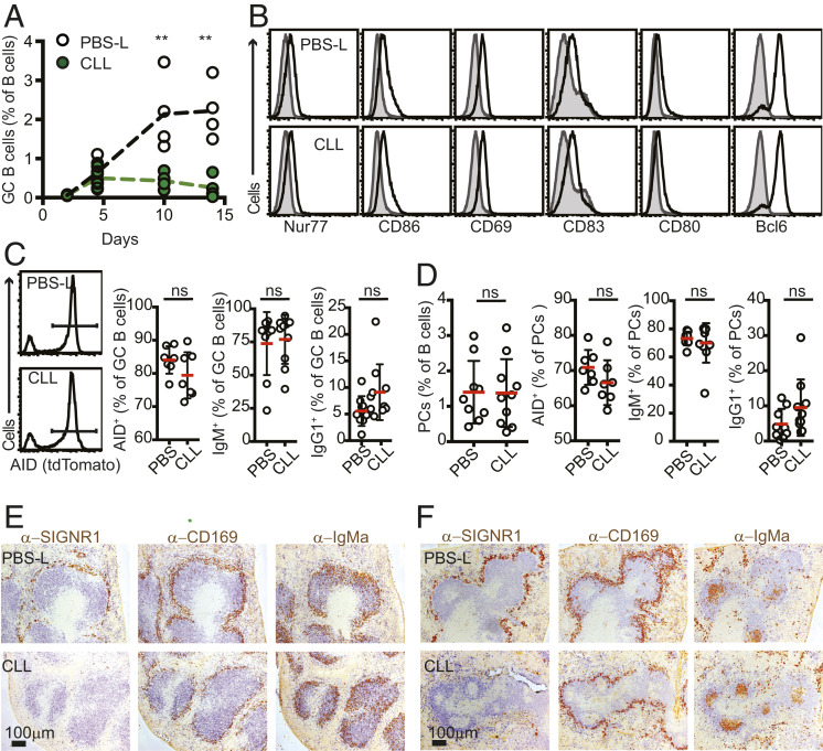
Early development of GC B cells is intact in SIGN-R1 macrophage-depleted mice. (A) CLL or PBS-L were injected at 3 wk before i.p. immunization with SRBCs. Spleens were analyzed by flow cytometry at the indicated time points to determine the frequency of GC B cells (B220+IgDlowCD95+GL7+ live cells) among total B cells. The experiment represents one out of two experiments performed (n ≥ 3). (B) Flow cytometry analysis of GC B cells from day 5 after SRBC immunization of mice treated with PBS-L (Top) or CLL (Lower) as above (n = 3). The gray histograms show staining in naïve B cells (B220+IgDhighCD95−GL7−) in immunized animals. Shown are the results of a representative experiment out of three independent experiments performed. (C and D) AID-tdTomato mice were treated and immunized as above with SRBCs. At 5 d postimmunization, spleens were analyzed. (C) Frequencies of dtTomato+, IgM+, and IgG1+ within the GC B cell pool (pregated on B220+IgDlowFAShighGL7high). The histograms show examples of tdTomato gating. (D) Frequencies of intracellular staining of IgM and IgG1 within the plasma cell (PC) pool. PCs were defined as B220lowCD138+ cells. Data in C and D are pooled from two independent experiments. (E and F) Distribution of HEL-specific activated B cells. Mice were treated with PBS-L or CLL and 3 wk later were cotransferred with HEL-specific CD45.1+ MD4 B cells and OTII T cells, followed by i.p. immunization with HEL-OVA. Spleens were collected and snap-frozen at 3 d (E) or 5 d (F) postimmunization. The distribution of activated B cells was assessed by IHC staining for IgD (to highlight naïve endogenous B cells; blue) and IgMa (to identify transferred MD4 B cells; brown). Selective ablation of SIGN-R1 macrophages, but not of CD169+ cells, was confirmed by staining consecutive sections for IgD (blue) and SIGN-R1 or CD169 (brown). The data are from one out of three independent experiments performed. In each experiment, sections from at least three spleens per group were analyzed.

