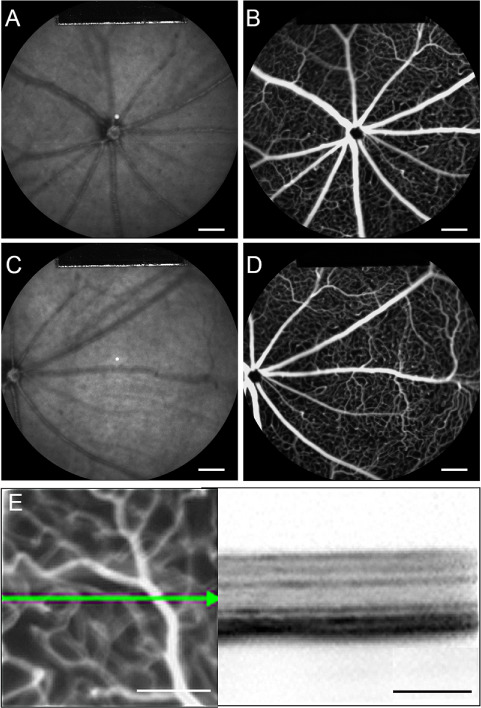
Figure 1.

Multimodal live images of retina from Pde6gcreERT2/+;Vhlflox/flox control mouse displays intact retinal vasculature. (A and C) Fundus IR imaging (800 nm) of the Pde6gcreERT2/+;Vhl flox/flox mouse model centered on the optic disc. (B and D) FFA of the fundus using fluorescein dye and a blue (488 nm) laser stimulus. (E) OCT cross section of Pde6gcreERT2/+;Vhl flox/flox using near-IR reflectance (820 nm) modalities. A and B are centered on the optic disc. C and D were used to observe the peripheral retina with the optic disc centered on the left. (white scale bar = 1 mm; black scale bar = 200 μm; green arrow = OCT scan)
