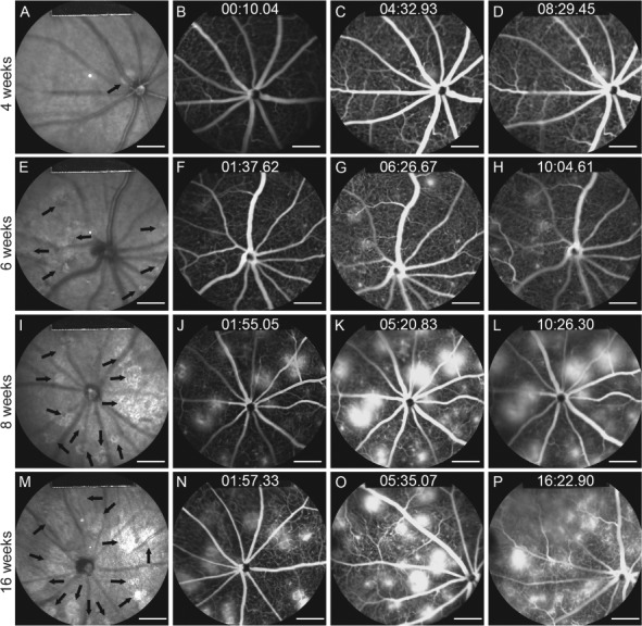
Figure 2.

IR imaging and fluorescein angiography show progressive neovascularization and leakage after HIF induction at postnatal day 7. Tamoxifen was administered to the Pde6gcreERT2/+;Vhlflox/flox mouse model at P7 for 3 days. (A, E, I and M) IR images of the fundus were collected at 4, 6, 8 and 16 weeks. Black arrows indicate hyporeflective lesion areas. FFA images were collected at early, middle and late phases at weeks 4 (B–D), 6 (F–H), 8 (J–L) and 16 (N–P) (scale bar = 1 mm).
