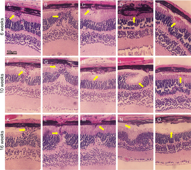
Figure 6.

Subretinal neovascularization, hemorrhage, exudation, retinal folds and exudative detachment after Vhl−/−. Tamoxifen was administered to Pde6gcreERT2/+;Vhlflox/flox mice at P7 for 3 days to generate Pde6gcreERT2/+;Vhl−/− mice. Eye cups were then harvested, and HE staining was performed to observe retinal structures at weeks 6 (A–E), 10 (F–J) and 16 (K–O) (yellow arrows = various retinal lesions; scale bar = 50 μm).
