Figure 8.

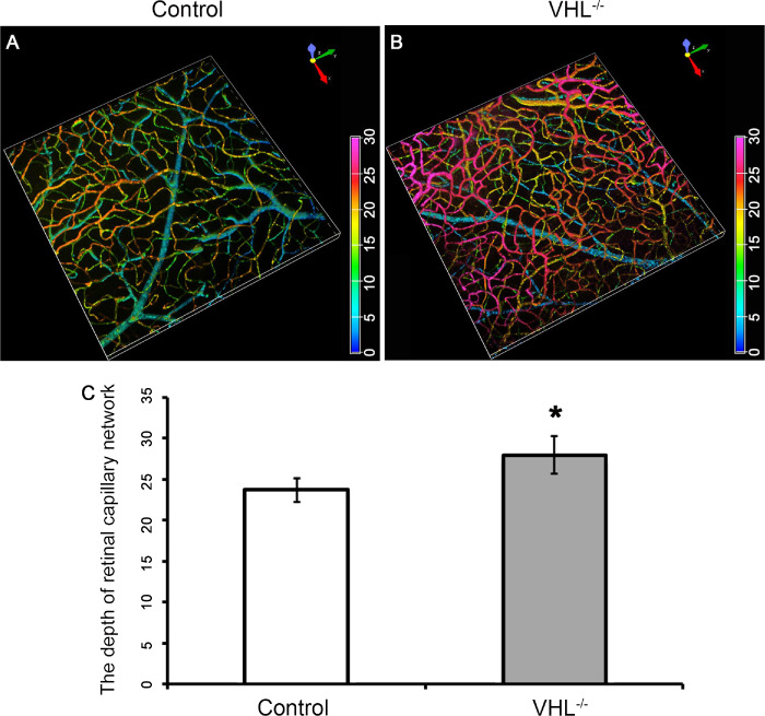
Increased capillary density after Vhl−/− in the Pde6gcreERT2/+;Vhl−/− mouse model. 3D remodeling of the vessels with immunofluorescence staining in the retinal flat mount in a Pde6gcreERT2/+;Vhlflox/flox (A) and Pde6gcreERT2/+;Vhl−/− (B) mouse at week 6. (C) Bar graphs quantifying the depth of retinal capillary network measured in the retinal flat mounts from control and Vhl−/− mice. Anti-CD31 was used as primary antibody (color spectrum = depth of blood vessels within the retina; *P < 0.05).
