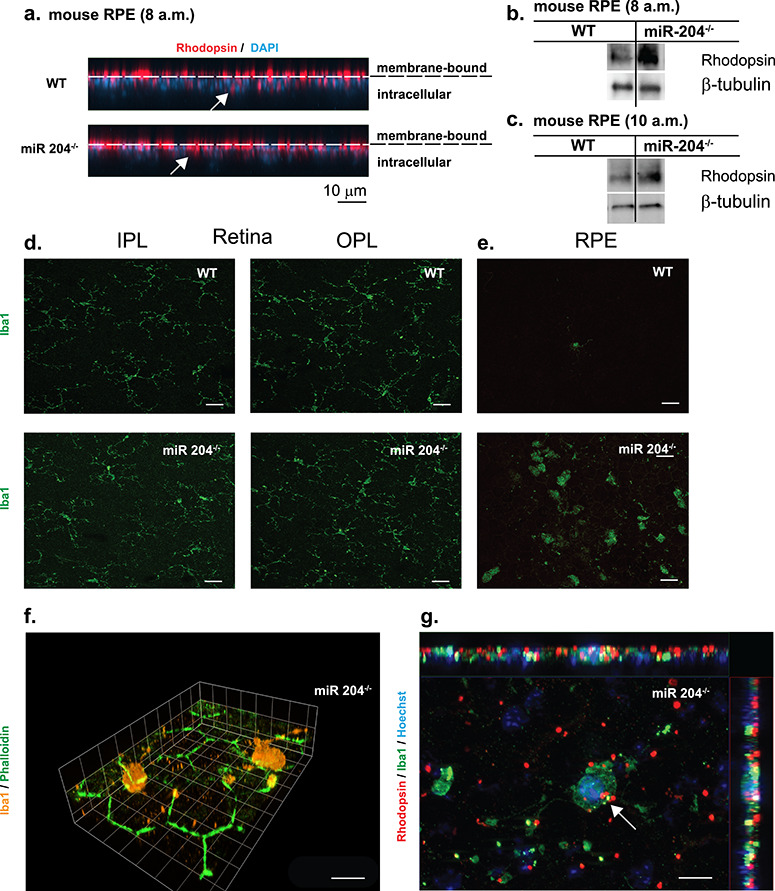
Figure 4.

Increased rhodopsin accumulation and microglial activation in miR-204−/− mice. (a) Representative confocal images (XZ) of RPE whole mount tissues of 8- and 9-month-old WT and miR-204−/− mice (collected at 8 a.m.) immunostained for rhodopsin (red) and counterstained with DAPI. Dashed lines indicate boundary between membrane bound rhodopsin and internalized/intracellular rhodopsin. Scale bar, 10 μm. (b and c) Rhodopsin levels in the RPE of WT and miR-204−/− mice were measured by western blot (n = 5 animals) at 8 a.m. or 10 a.m., respectively. (d) Resident microglia in the retina of wild-type and miR-204−/− mice. Iba1-labeled (green) microglial cells localized to IPL and OPL in WT and miR-204−/− mouse (8–9 months). Magnification, 40×. Scale bar, 20 μm. (e) Immunohistochemical staining for Iba1 (green) of RPE whole mount tissues from 8 to 9-month-old WT and miR-204−/− mice. Scale bar, 20 μm. (f) Iba1+ (orange) microglial cells are shown infiltrating the apical surface of the RPE in the miR-204−/− mice. Phalloidin staining (green) reveals the RPE’s regular hexagonal mosaic. Magnification, 100×. Scale bar, 10 μm. (g) Iba1+ (green) microglial cells on the apical surface of the RPE in miR-204−/− mice co-localized (indicated by the white arrow) with rhodopsin (red). Nuclei labeled with Hoechst (blue). Magnification, 100×. Scale bar, 10 μm.
