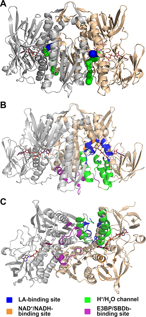
Figure 2.

Functionally important regions in hE3. (A) The hE3 structure (PDB ID: 6I4Q) is represented in a similar way, but in a different orientation, to Fig. 1 for reference to B and C (colors and all representations are as for Fig. 1). (B and C) Functionally important regions in hE3 under investigation in this study are represented in two orientations in monomer A. Selected residues from the adjacent monomer also contribute to the formation of the active site, the LA-binding site and the H+/H2O channel. These residues are not distinguished by coloring in this figure (see also Supplementary Material, Fig. S6).
