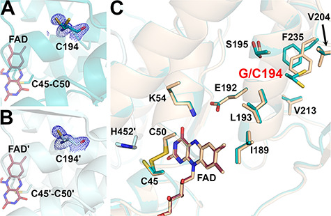
Figure 5.

Structure of the G194C-hE3 variant (PDB ID: 6I4P). (A and B) Cys194 is shown in (2mFo-DFc) composite omit map (2.0σ) in chains A and B, respectively. Based on indubitable difference densities, Cys194 could only be modeled with two alternative conformers in chain A (A). Active site disulfide and FAD (raspberry) are represented with sticks. (C) Active and substitution sites are shown in the superimposed structures of G194C-hE3 (dark teal; FAD: raspberry) and hE3 (beige; FAD: beige) in chain A. Cartoons are semitransparent and peptide segment 160–180 was removed for clarity.
