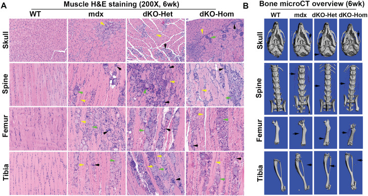
Figure 6.
Characterization of muscle and bone of 6-week-old mice. (A) H&E staining (200×) of different skeletal muscle tissue from 6-week-old WT, mdx, dKO-Het and dKO-Hom mice. Severe muscle histopathology is evidenced by centrally nucleated myofibers and inflammation, and heterotopic bone formation is shown by the presence of bony structures. Yellow, green and black arrows point to the centrally nucleated myofiber, heterotopic bone structure and inflammation, respectively. (B) MicroCT 3D overview of bone tissue at 6 weeks of age. Bone structures of the skull, spine, femur and tibia of mdx, dKO-Het and dKO-Hom mice were similar to those of WT control mice; however, heterotopic bone formation in the soft tissue surrounding the bone tissue (black arrows) was obvious in the mdx, dKO-Het and dKO-Hom mice.

