Figure 5.
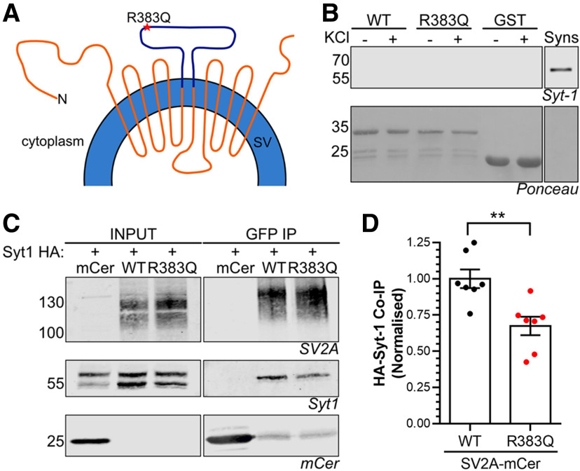
SV2A R383Q displays reduced binding to Syt1. A, Diagram depicts SV2A in the SV membrane with the cytoplasmic loop fused to GST highlighted in purple. B, Glutathione beads coupled to either GST alone, wild-type (WT) GST-SV2A356–447 or R383Q GST-SV2A356–447 were used to extract interaction partners from nerve terminal lysates which had previously been challenged with or without 30 mm KCl. A representative western blot is displayed for Syt1 in addition to Ponceau staining to reveal GST fusion proteins. Synaptosome lysates (Syns) were also present as a positive control (representative of five independent experiments). C, D, HA-tagged Syt1 was co-expressed with either mCerulean empty vector (mCer), WT SV2A-mCer or R383Q SV2A-mCer in HEK cells. Immunoprecipitation of mCer was performed from cell lysates. C, Representative western blots displaying the amount of SV2A, Syt1, or mCer present in either the input (2.5%) or immunoprecipitate. D, Quantification of the amount of HA-Syt1 co-immunoprecipitated (co-IP) was normalized to the amount of SV2A-mCer ±SEM (n = 7, Student's t test, **p = 0.0035).

