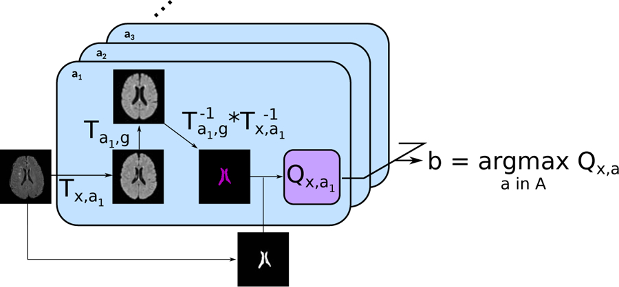
Figure 2: Principle of the proposed MAR framework.

For each subject, the input image was first registered to each of the atlases a ∈ A, which had been previously registered to the general atlas. The ventricles segmented on the general atlas Vg are then propagated first to each atlas a, and then to the subject’s image space. The propagated ventricles Vx,a,g were subsequently compared to VCNN, the subject’s ventricles segmented using the proposed automatic algorithm. Finally, the atlas maximizing the registration quality was selected for the intermediary registration step.
