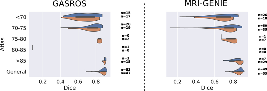
Figure 8: Comparison of multi-atlas registration using automated (blue) and manual (orange) segmentation of the ventricles in subject space.

The number of scans assigned to each atlas is indicated on the right of each plot for both automated and manual ventricle segmentations.
