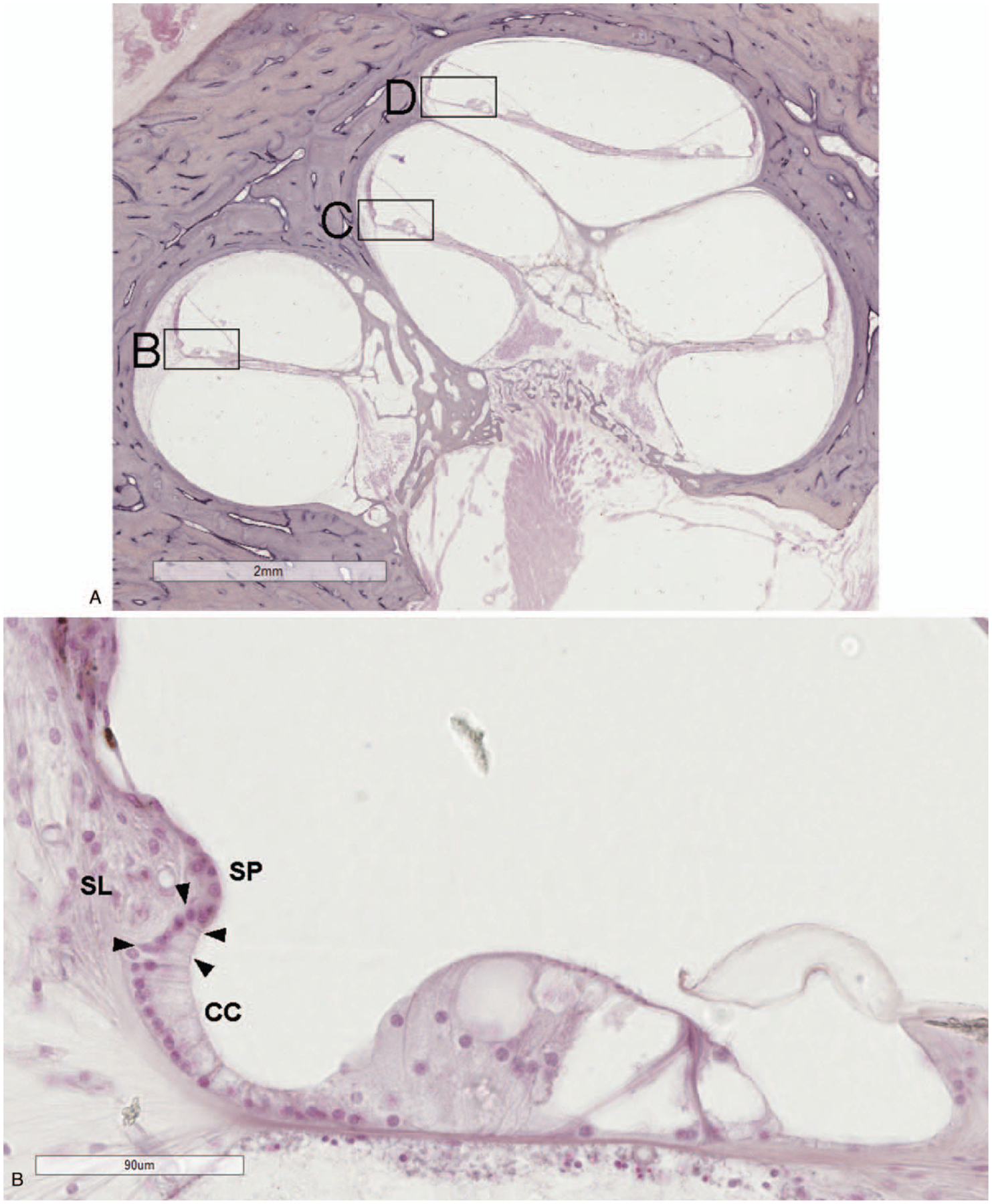
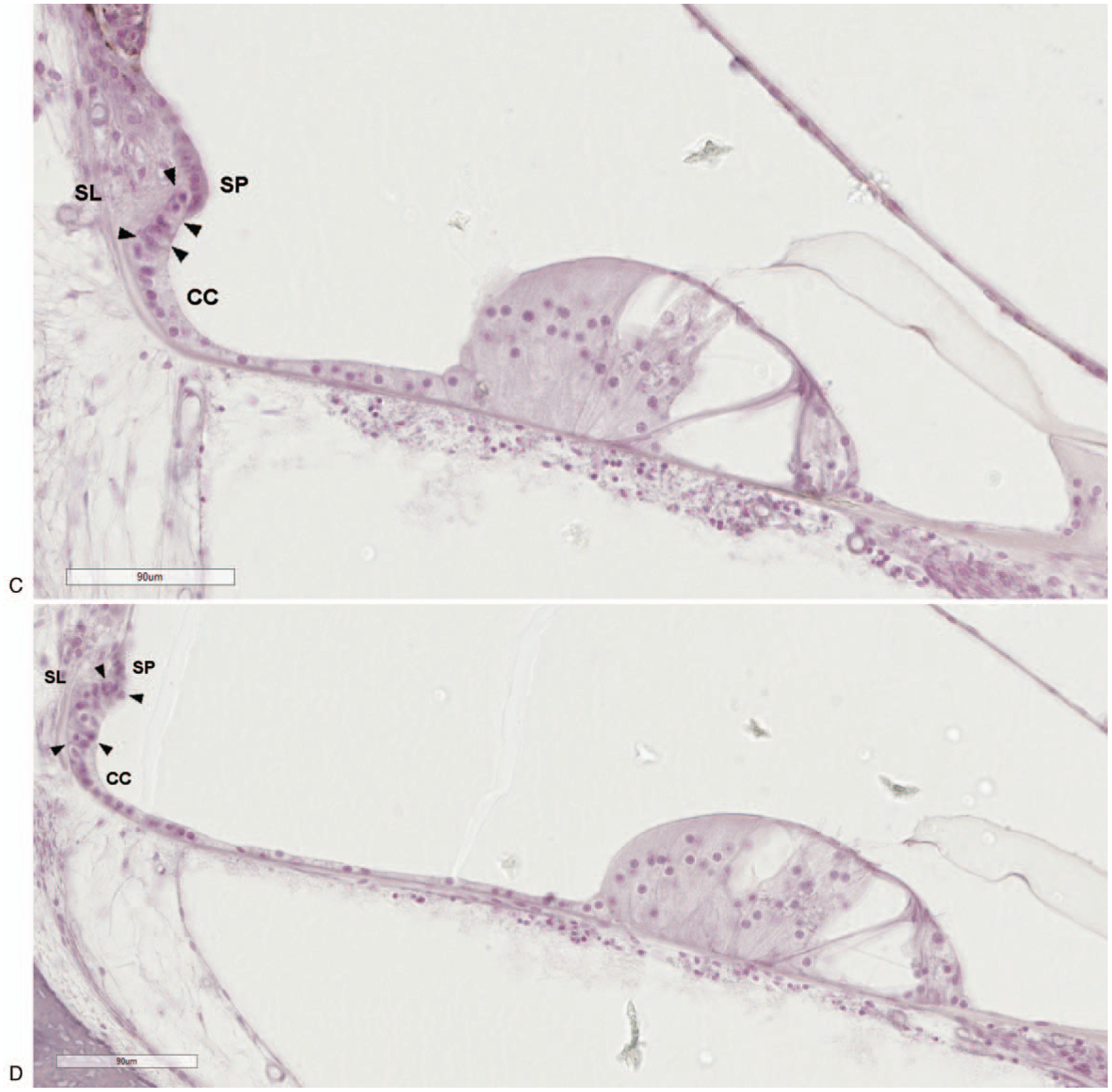
FIG. 1.


A, Light microscopy image of the human cochlea with boxes marking areas of high magnification. The outer sulcus region in the upper basal (B), upper middle (C), and upper apical (D) turns. The outer sulcus cells (arrowheads) are located between the last of the Claudius cells (CC) and the spiral prominence (SP). Their root-like processes extend into the adjacent spiral ligament (SL). (hematoxylin and eosin).
